Peptide · Checked
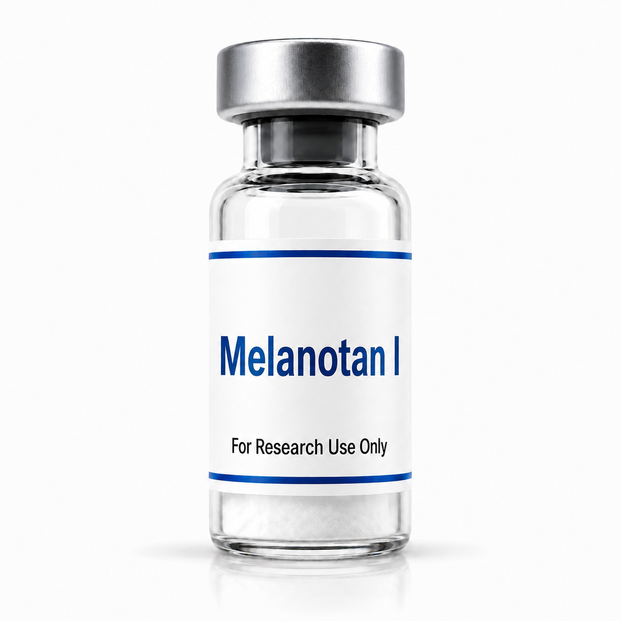
Melanotan I
Every price, divided down to the milligram.
Sticker prices hide what a compound actually costs, because vial sizes differ. So we divide the listing by its stated peptide content and show the real cost per milligram.
Vendors pay us a commission when you buy through our links. It never changes the order — rows are sorted by measured price per mg, and every flag is computed from the data shown. Read the full disclosure
Where to buy
One listing trackedHow this page is funded: vendors listed here pay us a commission when you buy through our links. Commissions never change the ranking — rows are ordered by measured price per mg, and flags are computed from the data on this page. Sponsored placements, if any, are always labeled.
On this page
How to read these rows
Verify the COA
A certificate of analysis should come from an independent lab, reference the exact batch number on the label, and be recent. Vendor-issued documents without a named third-party lab carry less weight — the COA row links to what each vendor publishes.
Match the format
Lyophilized vials and sprays are different products with different concentrations. That is why sprays sit outside the $/mg ranking — compare vials to vials, sprays to sprays.
Count the total cost
Shipping fees, free-shipping thresholds and active discount codes can flip which vendor is actually cheapest for your order size. The $/mg figure is the baseline; the shipping and code details finish the math.
How we rank
The row below carries a price per mg figure: listed price divided by stated peptide content, excluding shipping and codes. It is the same calculation applied on every compound page, so a figure here is directly comparable to one anywhere else on the site.
We currently track a single listing for this compound. When a second vendor is added the rows are ordered by measured $/mg, and the flags for testing and shipping are computed from the data shown. Each row carries its own verification date.
About Melanotan I
Names and development codes
Afamelanotide (INN) · SCENESSE (trade name) · NDP-α-MSH · NDP-MSH · Melanotan · Melanotan-1 · Melanotan I · MT-I · CUV1647 · EPT1647 · MBJ-05
Storage and handling
- Appearance
- white to off-white lyophilized powder
- Solubility
- water and aqueous laboratory buffers
- Storage, short-term
- 2–8 °C
- Storage, long-term
- −20 °C, desiccated, protected from light
Structure: two substitutions from native α-MSH
Melanotan I is native α-MSH with exactly two changes. Both are at positions that determine how long the peptide survives and how tightly it binds.
Position 4 Methionine → Norleucine (Nle)
Removes the oxidation-sensitive sulfur-containing residue. Norleucine is isosteric with methionine but carries no thioether, so the molecule is not subject to methionine oxidation.
Position 7 L-Phenylalanine → D-Phenylalanine
Stereochemical inversion at a single centre. The D-configuration resists proteolytic cleavage and is associated in the structure-activity literature with substantially increased potency.
Everything else — the N-terminal acetyl group, the C-terminal valinamide, and the remaining eleven residues — is unchanged from α-MSH.
Note on relative mass: α-MSH has molecular formula C77H109N21O19S and a molecular weight of 1664.88 g/mol. Melanotan I is the LIGHTER of the two at 1646.87 g/mol, because replacing methionine's sulfur with a methylene group in norleucine removes more mass than it adds. This is counterintuitive if one assumes a modified analog must be heavier.
Active site
The His-Phe-Arg-Trp motif at residues 6-9 is the conserved melanocortin recognition element, retained in Melanotan I with the phenylalanine in D-configuration.
Reported potency
Sources describe Melanotan I as approximately 1,000-fold more potent than native α-MSH in stimulating melanogenesis, attributed to the combination of increased receptor affinity and resistance to enzymatic degradation.
Receptor pharmacology
Melanotan I is classified in the FDA label and the primary literature as a melanocortin 1 receptor (MC1R) agonist. As a close analog of α-MSH, which itself signals across the melanocortin receptor family, it retains activity at other melanocortin receptor subtypes. Sources describe it as MC1R-preferring rather than strictly MC1R-exclusive, and some characterize it as a non-selective melanocortin agonist with highest affinity at MC1R.
That distinction matters when comparing it to Melanotan II. The practical difference between the two is not that one is selective and the other is not — it is that Melanotan I is used predominantly in MC1R-centred pigmentation and photobiology work, while Melanotan II is studied across broader melanocortin receptor activity.
Signaling cascade
MC1R is a Gs protein-coupled receptor expressed on melanocytes.
Agonist binding activates adenylyl cyclase, raising intracellular cyclic AMP. cAMP-dependent signaling upregulates microphthalmia- associated transcription factor (MITF), which in turn drives expression of tyrosinase and related melanogenic enzymes. The pathway shifts pigment synthesis toward eumelanin rather than pheomelanin.
Melanogenesis and pigmentation
The core body of work. MC1R activation initiating cAMP-dependent upregulation of tyrosinase and melanogenic enzyme expression, with the eumelanin/pheomelanin balance as the measured endpoint.
DNA repair
A distinct and less widely reported line of research. MC1R activation has been reported to enhance nucleotide excision repair through cAMP-dependent signaling, with protein kinase A phosphorylating ATR at serine 435 and improving XPA recruitment to sites of DNA damage. Increased expression of base excision repair enzymes including OGG1 and APE1 has also been reported. The functional readout in these models is accelerated clearance of UV-induced photolesions.
Photobiology and UV response
Studies examining the relationship between melanocortin receptor activity, pigment-cell signaling, and ultraviolet response mechanisms in melanocyte models.
Erythropoietic protoporphyria
The approved indication. Randomized controlled trials measured pain-free sunlight exposure duration, with reported medians of approximately 64 hours in the afamelanotide arm versus approximately 40 hours on placebo (Langendonk et al., NEJM 2015).
Anti-inflammatory signaling
Melanocortin receptor engagement on macrophages has been reported to modulate NF-κB signaling, with MC1R and MC3R activation reducing pro-inflammatory cytokine production and nitric oxide generation, and increasing IL-10.
Oxidative stress and neuroprotection
MC1R signaling has been examined in relation to reactive oxygen species handling and antioxidant enzyme expression, including superoxide dismutase, glutathione peroxidase, and catalase, via PI3K/Akt/Nrf2 signaling. Separate work has examined AMPK, SIRT1, and PGC-1α pathways in models of cerebral injury.
Hepatic models
Melanocortin signaling has been examined in endotoxin-induced liver inflammation models, with reported reductions in hepatic neutrophil infiltration.
Vitiligo
Afamelanotide in combination with narrow-band UVB has been examined in nonsegmental vitiligo.
Origin
Developed from work at the University of Arizona, licensed via Competitive Technologies to the Australian company Epitan, which subsequently became Clinuvel Pharmaceuticals. The development code CUV1647 dates from that period.
Approval
Approved by the FDA in October 2019 and by the EMA, marketed as SCENESSE, for the prevention of phototoxicity and reduction of light-exposure pain in adults with erythropoietic protoporphyria.
Legal status
Prescription-only in the United States, European Union, and United Kingdom. Schedule 4 (prescription only) in Australia.
Route of administration: subcutaneous implant.
Research material
Material supplied for laboratory use is a research chemical, not the approved pharmaceutical product. It is not a drug, food, cosmetic, or dietary supplement as supplied, has not been evaluated by the FDA in that form, and is not intended for human or veterinary use.
Structure linear 13-residue cyclic 7-residue linear lactam 13-residue CAS 75921-69-6 121062-08-6 581-05-5 Formula C78H111N21O19 C50H69N15O9 C77H109N21O19S Molecular weight 1646.87 g/mol 1024.2 g/mol 1664.88 g/mol PubChem CID 16154396 / 16197727 92432 16133793 Receptor profile MC1R-preferring non-selective endogenous MC1R/3R/4R/5R ligand Regulatory status FDA and EMA never submitted endogenous approved (2019) anywhere peptide Approved use erythropoietic none n/a protoporphyria
Melanotan I and Melanotan II are frequently conflated in secondary sources despite differing in ring structure, residue count, molecular weight by more than 600 Da, receptor profile, and regulatory standing. Literature on one does not transfer to the other.
Specifications
| Attribute | Value |
|---|---|
| Molecular formula | C78H111N21O19 |
| Molecular weight | 1646.87 g/mol |
| CAS Number | 75921-69-6 |
| PubChem CID | 16154396 · 16197727 |
| InChIKey | UAHFGYDRQSXQEB-LEBBXHLNSA-N |
| ChemSpider | 17310725 |
| DrugBank | DB04931 |
| UNII | QW68W3J66U |
| KEGG | D10511 (acetate salt: D11334) |
| ChEBI | CHEBI:136034 |
| ChEMBL | ChEMBL441738 |
| IUPHAR/BPS | 1324 |
| EPA CompTox | DTXSID40226843 |
| ATC code | D02BB02 |
| Residue count | 13 |
| Structure class | linear synthetic tridecapeptide |
| Structural shorthand | [Nle4, D-Phe7]-α-MSH |